-
- Pathogenic Mechanism of Stroke
- Exploration of the Role of Differential Targets in StrokeExploration of the Role of Cell Death in StrokeExploration of the Role of Different Cell Types in StrokeExploration of the Role of Other Components in Stroke
- Animal Modeling of Stroke
- Animal Modeling of Ischemic StrokeAnimal Modeling of Hemorrhagic StrokeIn Vitro Modeling of Stroke
- In Vitro Modeling of Ischemic Stroke
- In Vitro Modeling of Hemorrhagic Stroke
-
Custom Endovascular Filament Model of Middle Cerebral Artery Occlusion (MCAO)
The middle cerebral artery (MCA) and its branches are the cerebral vessels most severely affected by ischemic stroke, accounting for approximately 70% of infarctions. Thus, occlusion of this artery most closely resembles focal cerebral ischemia stroke in humans. The most common method of focal ischemic stroke is intraluminal thread occlusion of the MCA, which has been used in more than 40% of stroke research. The endovascular filament MCAO modeling involves surgical filament insertion of the external carotid artery threaded into the internal carotid artery (ICA) until prompted by obstruction of the MCA at the origin, in the cessation of blood flow and subsequent cerebral infarction. The suture is located at the junction between the anterior cerebral artery and the middle cerebral artery and is retained in place for a predetermined period or permanently.
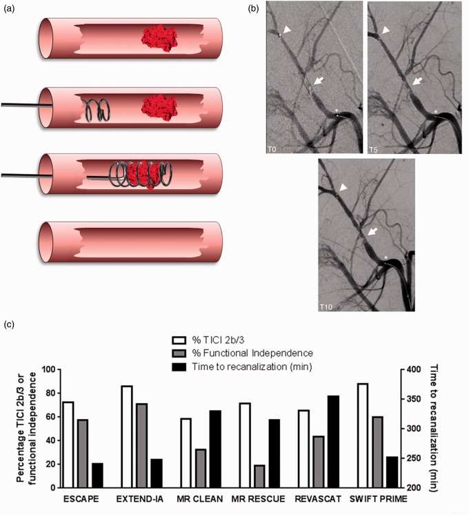 Fig. 1. Illustration of endovascular thrombectomy after acute ischemic stroke. (Sutherland et al., 2016)
 Custom Endovascular Filament Model of MCAO
Ace Therapeutics is proud to provide customized intraluminal filament model of MCAO to researchers in the stroke field. Ace Therapeutics has a wealth of experience and a dedicated team of scientists that allow us to model permanent or transient MCAO (pMCAO or tMCAO) focal ischemic stroke according to the specific needs of our clients. All studies can be customized and adapted to meet customer needs.
Animal Modeling Method
We can construct intraluminal filament model of MCAO in animals. The approximate procedure is as follows:
1) Under the operating microscope, the bifurcation of the right common carotid artery is exposed through a midline incision in the neck.
2) A 4-0 monofilament nylon suture with its tip rounded near a flame is introduced into the right external carotid artery and advanced into the internal carotid artery for a length of 17~20 mm from the bifurcation.
3) These methods place the tip of the suture at the origin of the anterior cerebral artery, thereby occluding the middle cerebral artery. The suture is left in place until death. If removal is followed by suturing at certain time intervals (30 minutes, 1 hour, or 2 hours), reperfusion (transient ischemia) is achieved; if the filament is left in place (24 hours) the procedure is suitable as a model of permanent ischemia.
4) Following MCA occlusion, animals are allowed to awaken from anesthesia. Surgical mortality is <10% in this model.
Animal Model Characterization and Validation
To assess the severity of cerebral infarction, we can stain brain sections with 2,3,5-triphenyl tetrazolium chloride (TTC) to determine ischemia in brain tissue. We also offer cognitive/behavioral tests to measure spatial memory, contralateral motor function and coordination (stairs, adhesion removal test, water maze, grip test).
Advantages of Our Custom Endovascular Filament Model of MCAO
- Our customized endovascular filament model of MCAO has been carefully designed to provide high reproducibility
- With a customized endovascular filament model of MCAO, researchers can accurately manipulate occlusion times and are able to explore the effects of different ischemia durations on brain tissue and functional outcomes
- The presence of significant ischemic semi-dark bands in the customized intraluminal filament MACO model allows researchers to identify affected areas of the brain and assess the extent of ischemic damage
Applications of Endovascular Filament Model of MCAO
- For stroke preclinical neuroprotective drug studies
- Study the neuroplastic rearrangements and neuro-regenerative cues after stroke
- Characterization of stroke-related genes and proteins
At Ace Therapeutics, our endovascular filament model of MCAO provides a more comprehensive approach for your focal cerebral ischemic stroke preclinical research. Need to know more? Contact us and let one of our experts provide you with all the answers you need.
Reference- Sutherland, B. A., et al. (2016). The transient intraluminal filament middle cerebral artery occlusion model as a model of endovascular thrombectomy in stroke. Journal of Cerebral Blood Flow & Metabolism, 36(2), 363-369.
All of our services are intended for preclinical research use only and cannot be used to diagnose, treat or manage patients.
0Inquiry Basket