-
- Pathogenic Mechanism of Stroke
- Exploration of the Role of Differential Targets in StrokeExploration of the Role of Cell Death in StrokeExploration of the Role of Different Cell Types in StrokeExploration of the Role of Other Components in Stroke
- Animal Modeling of Stroke
- Animal Modeling of Ischemic StrokeAnimal Modeling of Hemorrhagic StrokeIn Vitro Modeling of Stroke
- In Vitro Modeling of Ischemic Stroke
- In Vitro Modeling of Hemorrhagic Stroke
-
Custom Middle Cerebral Artery Occlusion (MCAO) Model by Transcranial Electrocoagulation
A major possibility for inducing ischemic stroke is occlusion of the more terminal vessels supplying the brain, and transcranial occlusion is a reliable option. A craniectomy requires opening the skull and incising the dura mater to directly block the proximal cerebral arteries. There are two main methods to build the model, including occlusion of the proximal middle cerebral artery (MCA) alone by direct electrocoagulation, ligation, transection, and photothrombosis, and combined occlusion of the MCA and bilateral common carotid artery (three-vessel occlusion model, 3-VO model) or ipsilateral common carotid artery. This technique allows for both permanent and temporary occlusion, depending on the duration of the occlusion.
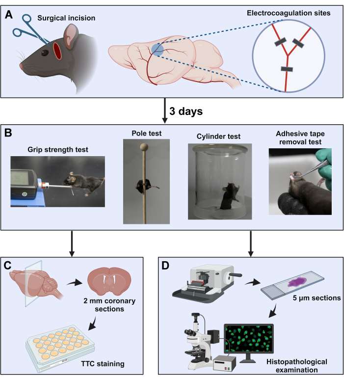 Fig. 1. Induction of acute ischemic stroke in mice using the distal middle artery occlusion technique. (Leng et al., 2023)
Custom MCAO Model by Transcranial Electrocoagulation
Ace Therapeutics is a leading company specializing in stroke science, providing customizable the permanent distal middle cerebral artery occlusion (MCAO) model by transcranial electrocoagulation in animals for ischemic stroke research. Our team of highly skilled experts with extensive industry experience combines extensive neuroscience knowledge with cutting-edge technology to provide superior support for preclinical stroke research.
Animal Modeling Method
Our transcranial occlusion modeling uses electrocoagulation to occlude the MCA, resulting in permanent occlusion. Electrocoagulation is a craniotomy that exposes the middle cerebral artery and cauterizes the corresponding area of the middle cerebral artery with an electrode, blocking the blood vessel and causing cortical or subcortical infarction in the area of the middle cerebral artery's blood supply. Our experts have modified the traditional electrocoagulation approach to create a more stable and reproducible mouse model of focal cerebral ischemia.
Animal Model Characterization and Validation
We offer multiple measures to analyze stroke outcomes, such as laser scatter measurements, magnetic resonance imaging, behavioral testing, or histological analysis. In this model, we can perform infarct volume measurements using 2,3,5-triphenyl tetrazolium chloride (TTC)-stained serial coronal brain sections 7 days after stroke induction. In addition, we can assess behavioral deficits after MCA coagulation by analyzing the anterior claw using an asymmetric cylindrical test.
Advantages of Our Custom MCAO Model by Transcranial Electrocoagulation
- Our customized MCAO model by transcranial electrocoagulation can induce precise and reproducible occlusion of target cerebral arteries
- Our customized approach incorporates real-time visualization techniques that allow researchers to visually confirm successful occlusion of the MCA
- We have the freedom to modify occlusion parameters such as duration and severity to model different stroke subtypes or to study specific problems
Applications
- Study the reperfusion support neuronal survival, rescue of vascular phenotype, and functional recovery after stroke
- Evaluate ischemic stroke drug efficacy
At Ace Therapeutics, our customizable permanent distal MCAO model by transcranial electrocoagulation provides a more comprehensive approach for your preclinical ischemic stroke research. Need to know more? Contact us and let one of our experts provide you with all the answers you need.
Reference- Leng, C., Li, Y., Sun, Y., et al. (2023). Induction of Acute Ischemic Stroke in Mice Using the Distal Middle Artery Occlusion Technique. JoVE (Journal of Visualized Experiments), (202), e66134.
All of our services are intended for preclinical research use only and cannot be used to diagnose, treat or manage patients.
0Inquiry Basket